The research results of Professor Zeng Yuan-shan's Team at Zhongshan School of Medicine have been published in Biomaterials
Source: Zhongshan School of Medicine
Written by: Zhongshan School of Medicine
Edited by: Tan Rongyu, Wang Dongmei
Is central nerve regeneration still difficult in adult mammals? Recently, a team led by Professor Zeng Yuan-shan at Zhongshan School of Medicine of Sun Yat-sen University found that altering the extracellular matrix (ECM) of the optic nerve can reverse its inhibiting tissue microenvironment of nerve regeneration.
Compared with peripheral nerve regeneration, regeneration after central nerve injury involves a series of more complex microenvironment and intracellular signal regulation mechanisms. How to optimize the microenvironment of injured tissue and improve the biomimetic structure of nerve graft is the main strategy and urgent problem to repair the central nerve injury by using the principle of tissue engineering. Autologous peripheral nerve or decellularized peripheral nerve grafts have been used to repair peripheral nerves because of their biomimetic structure and low immunogenicity, and have achieved well curative effects. In theory, central nerve grafts also have good structural bionic properties. However, they are rarely used as natural biomaterials for repairing nerve injury due to their strong inhibitory microenvironment of nerve regeneration. Therefore, optimization of the central nervous tissue microenvironment can provide a new idea for tissue-engineered nerve grafts to repair central nerve injuries.
On August 10, 2020, the team of Professor Zeng Yuan-shan published the research results online in the international TOP publication
Biomaterials (CAS Q1, IF = 10.273): Decellularization Optimizes the Inhibitory Microenvironment of the Optic Nerve to Support Neurite Growth. The co-authors are Sun Jia-hui (PhD student of Zhongshan School of Medicine) and Dr. Li Ge (associate researcher of Zhongshan School of Medicine); Professor Zeng Yuan-shan is the corresponding author.
The present study found that decellularization reversed the microenvironment of adult porcine optic nerve (ON) inhibiting nerve regeneration, made it more similar to the ECM of embryonic porcine optic nerve, optimized the function of optic nerve ECM, and made it support the straight growth of dorsal root ganglion (DRG) neurites. The distance of neurite growth on the decellularized optic nerve (DON) longitudinal-thikness slices was significantly longer than that on the normal ON. Compared to the ON, the neurite branches growing on the DON also increased significantly. Proteomics technology was used to analyze the ECM components of adult porcine ON, adult porcine DON and embryonic porcine ON. It was found that the protein components of the DON had a tendency to turn to embryo. The decellularization technique selectively removes some molecules that inhibit neurite growth, such as myelin-associated glycoproteins (MAG) and chondroitin sulfate proteoglycans (CSPGs) and remains the proteins that support neurite growth, including collagen Ⅳ (COL4) and laminin (LAM), etc. The COL4 and LAM, located on the connective tissue septum of DON longitudinal-thikness slices, have been showed to bind to integrin α1 (ITGA1, their receptor) on the DRG neurites, which is one of the factors leading to the straight growth of DRG neurites. This study provides a feasible techical path for optimizing functional transformation of the central nervous tissue microenvironment to be conducive to nerve regeneration, and also lays a theoretical foundation for the application of DON as a natural biological scaffold in nerve injury repair.
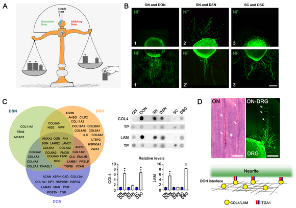
Note: A.) Schematic diagram showing the stable microenvironment of central nervous tissue was changed by the decellularization technique. The significant change in the ECM of central nervous tissue after decellularization is to decrease the components inhibiting nerve regeneration and increase proportion of the components promoting nerve regeneration, and contribute to the transformation of the microenvironment from steady state to stimulative state. B.) Showing the three nervous tissues (optic nerve, ON; sciatic nerve, SN; spinal cord, SC) and their corresponding decellularized nerve tissue (DON; DSN; DSC) on DRG neurite growth. C.) Wenn diagram showing the ECM protein components retained by three kinds of decellularized nervous tissues (DON, DSN and DSC). D.) Appearing the relationship between the connective tissue septum (white arrows) of normal optic nerve longitudinal-thikness slice and its supporting the straight growth of DRG neurites (white arrowheads). In addition, the pattern diagram shows the relationship between COL4/LAM on the DON interface and their receptor ITGA1 on the neurite.
This research was supported by grants from the National Natural Science Foundation of China, the National Key R&D Program of China, the Foundation of Guangdong Province and the Start-up Foundation of Guangdong Province.
Paper link:
https://www.sciencedirect.com/science/article/pii/S0142961220305354