One artificial intelligence system for peritoneal carcinomatosis has been developed and published in top surgical journal from colorectal surgery of the Sixth Affiliated Hospital of Sun Yat-sen University
Source: The Sixth Affiliated Hospital
Written by: The Sixth Affiliated Hospital
Edited by: Tan Rongyu, Wang Dongmei
Peritoneal carcinomatosis (PC) is considered to be the terminal stage of colorectal cancer (CRC) and obtains poor prognosis. Currently, the imaging tools for detecting PC is limited by low sensitivity, especially for these small PC nodes < 5mm in size. recently, the researchers in colorectal surgery in the sixth affiliated hospital of sun yat-sen university (sysu) of china has developed the first artificial intelligence (ai) system for diagnosis of pc in cooperation with tencent ai lab in shenzhen. the original article was published in the top surgical journal
Annals of Surgery (Impact factor: 10.13 points) with the title “Development and Validation of an Image-based Deep Learning Algorithm for Detection of Synchronous Peritoneal Carcinomatosis in Colorectal Cancer”. Dr. Zixu Yuan from the Sixth Affiliated Hospital of SYSU is the first author, and Professor Hui Wang is the corresponding author. Vice chief doctor Jian Cai, Dr. Wuteng Cao in radiology, and Dr. Yebiao Zhao have made critical attributions to this article.
Figure 1. AI system based on deep learning algorithm.
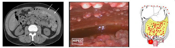
Figure 2. CT imaging of peritoneal carcinomatosis and extensive PC nodes in the abdominal wall.
CRC with synchronous PC was presented with the incidence of 5-10%, and PC incidence increased to 25-44% in recurrent cases with CRC. Professor Hui Wang said, “If PC is detected in early stage, patients can receive complete cytoreductive surgery (CRS) and the survival can be greatly prolonged”. Since 2018, the team has established cooperation with Tencent AI lab to create a ResNet3D system with convolutional neural network (CNN). This is the first AI system to diagnose PC in the world. It can recognize the features of primary tumor and also delineate radiological features of adjacent peritoneum and create one support vector model (SVM) classifier by AI algorithm. A total of 19,814 CT images were enrolled in the training set, and 7,837 CT images were enrolled in the test set.
The results show ResNet3D system spent only 34s to recognize and diagnose all of test images. The accuracy of ResNet3D + SVM classifier in diagnosis of PC was 94% (AUC:0.922), with both sensitivity and specificity of 94%. The performance of AI system was greatly superior to radiologists.
What is the medical clinical value? Dr. Zixu Yuan said, “The AI system we developed is a non-invasive novel detection system. Enhanced CT scans are widely used for diagnosis of abdominal tumors. This AI system can not only automatically recognize primary tumor, but also integrate features of adjacent peritoneum. It obtains high values in clinical applications. It will provide clinical reference for clinicians to make surgical plans, and provide evidence of optical treatment for CRC patients.”
Dr. Zixu Yuan said, this AI system can recognize radiological images in other hospitals and it will be transplanted to other medical center in the future. Through larger external validation cohort, clinical application of this AI system can be tested. It will help to resolve the difficult issue of PC diagnosis across the world.
Link to the paper:
https://pubmed.ncbi.nlm.nih.gov/32694449/