Source: Zhongshan Ophthalmic Center
Edited by: Zheng Longfei, Wang Dongmei
The team of Professor Jin Ma from Zhongshan Ophthalmic Center at Sun Yat-sen University published a research article entitled "Long-term continuous assessment of internal limiting membrane filling induced super-large macular hole healing" in
American Journal of Ophthalmology on April 2nd, 2022. The research team has for the first time introduced a modified internal limiting membrane filling technique in the treatment of super-large macular hole and achieved a satisfactory result. Through this long-term prospective study, the research team has revealed the characteristic closure pattern and visual outcome of super-large macular hole after internal limiting membrane filling, which has clarified the value and applicability of the modified internal limiting membrane flap filling technique in the treatment of super-large macular hole.
The super-large macular hole of diameter around or over 1000μm is a refractory macular disease and the surgery treatment is very challenging for the difficulty in macular hole closure. Previous attempts and explorations of surgical improvement (including lens capsule or amniotic membrane implantation) was limited for the unsatisfied healing state after surgery. The autologous internal limiting membrane flap should be the ideal material for implantation in closing the super-large macular hole. However, the application of internal limiting membrane filling has been controversial for the potential iatrogenic injury (RPE injury at the bottom of the macular hole) by the manual filling procedure as well as the debatable scar healing postoperatively. After six years of research, the team of Professor Jin Ma has successfully applied a modified internal limiting membrane flap filling technique in the treatment of super-large macular hole. The internal limiting membrane flap was naturally filled in the macular hole with the help of the surface tension of air during the air-fluid exchange procedure and minimized the risk of iatrogenic injury comparing to the conventional manual procedure. Over 90% of the cases has achieved macular hole close within postoperative one week even without any gas tamponade (with air tamponade only).
During the six-year’s research, the team of Professor Jin Ma has performed the modified internal limiting membrane filling technique in 97 cases of super-large macular hole, and a 15-months continuous follow-up was conducted to observe the anatomical and functional healing pattern. The team first summarized the closure pattern of super-large macular hole based on the OCT feature:
• Type I: Reconstruction of the foveal layered retinal structure within the outer limiting membrane (OLM), without any hyper/hypo-reflective mass perpendicular to the retinal structure (Ia: the OLM is continuous; Ib: the OLM is not continuous).
• Type II: Presence of hyperreflective mass perpendicular to the foveal retina structure, without any cysts within the foveal region (IIa: partially continuous with the foveal layered retina; IIb: discontinuous with all foveal layered retinas).
• Type III: Presence of cyst within the foveal retina structure, discontinuous with all foveal layered retinas.
The team also found that the macular hole closure pattern improved continuously and differed significantly in three groups (Fisher’s exact test, p<0 .05) (group a, hole diameter of 900–1,000 μm; group b, hole diameter of 1,000–1,100 μm; and group c, hole diameter of over 1,100 μm). type i closure pattern was probably attained in cases of hole diameter less than 1,000 μm, and for those of hole diameter over 1,000 μm, type ii closure pattern was most commonly seen, with a few type iii closure pattern appeared.

Figure 1 The pre- and post-operative fundus photography and OCT of a patient of super-large macular hole with modified internal limiting membrane filling.
The research has demonstrated that the macular hole diameter, macular sensitivity and fixation stability were significant predictors of postoperative best corrected visual acuity. The research has also proved that the filling of internal limiting membrane would not enlarge the central scotoma or affect the recovery of visual function, which clarified the value and applicability of the modified internal limiting membrane flap filling technique in the treatment of super-large macular hole.
Figure 2 MP-3 images of a patient with super-large macular hole before and 1 week after operation.
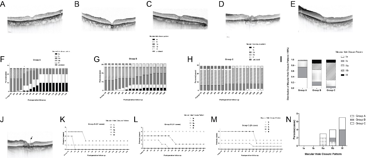
Figure 3 Continuous assessment of the anatomical change of super-large FTMH after ILM filling during the 15-month follow-up.
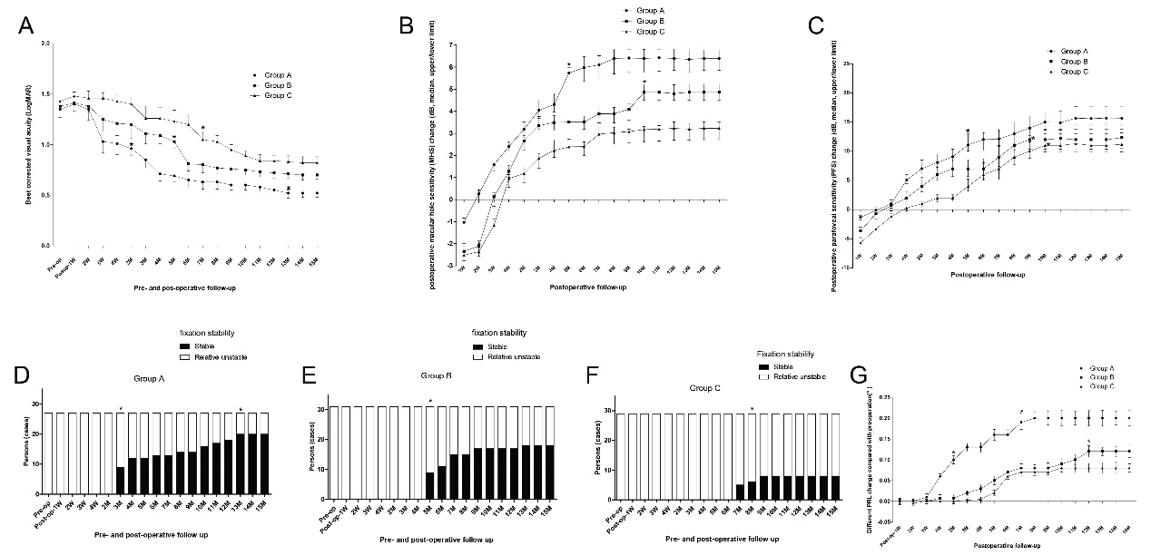
Figure 4 Continuous assessment of the visual function change of super-large FTMH after ILM filling during the 15-month follow-up
In summary, the team of Professor Jin Ma has first revealed the characteristic closure pattern and visual outcome of super-large macular hole after modified internal limiting membrane filling, and clarified the significance and value of this surgical technique. It has opened a new page for the surgical treatment of super-large macular hole. This technique is safer and easily extended, which may be preferable in the treatment of super-large macular hole.
Professor Jin Ma (from Zhongshan Ophthalmic Center at Sun Yat-sen University) is the corresponding author. Dr. Xiling Yu (from Zhongshan Ophthalmic Center at Sun Yat-sen University) is the first author. This work was supported by grants from the National Natural Science Foundation of Guangdong, China (2020A1515010190) and the Science and Technology Program of Guangzhou, China (202102010274). Thank Prof. Yizhi Liu, Prof. Ling Jin, and Prof. Xiang Chen (from Zhongshan Ophthalmic Center at Sun Yat-sen University) for the support and help of this research.
Link to the article:
https://www.ajo.com/article/S0002-9394(22)00133-7/